Recently I have been able to spend a little time Staffordshire University’s environmental scanning electron microscope (ESEM). I’d like to share some of the images that were captured.
Click any of the images for the full sized version!
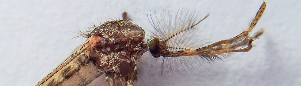
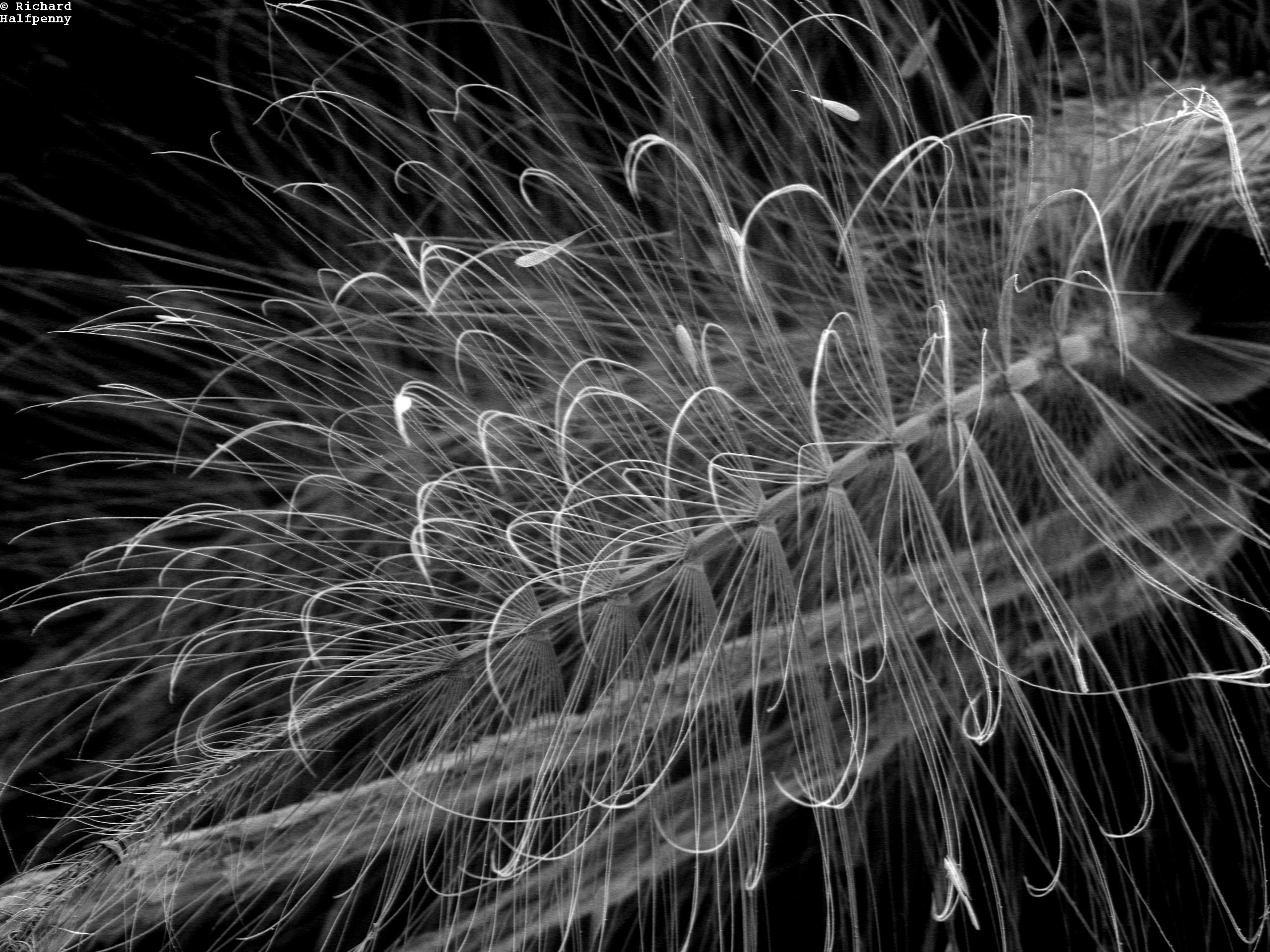
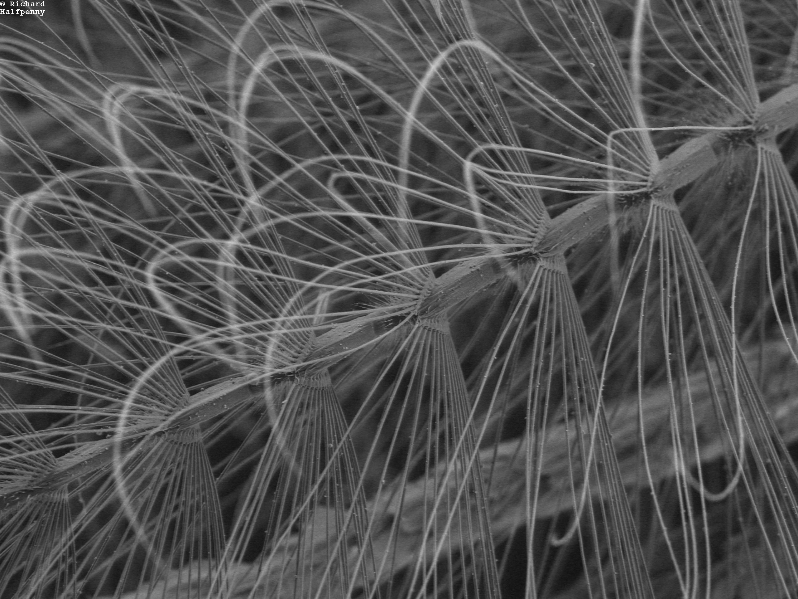
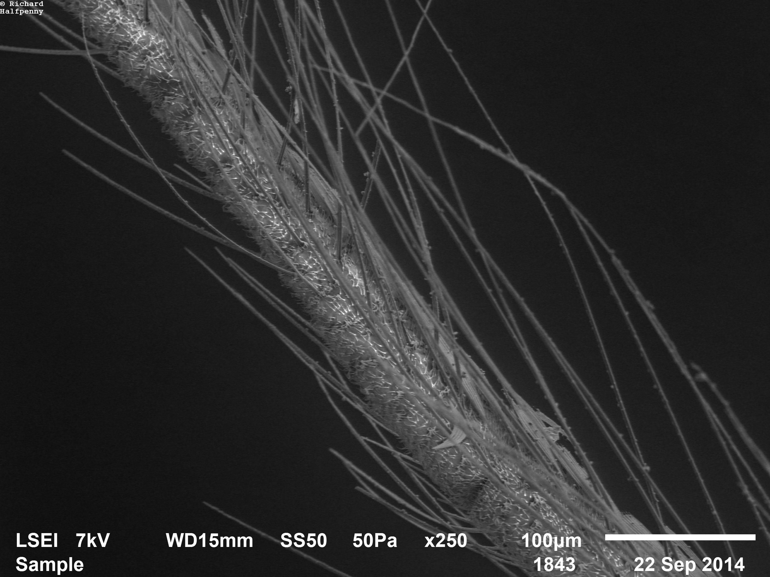
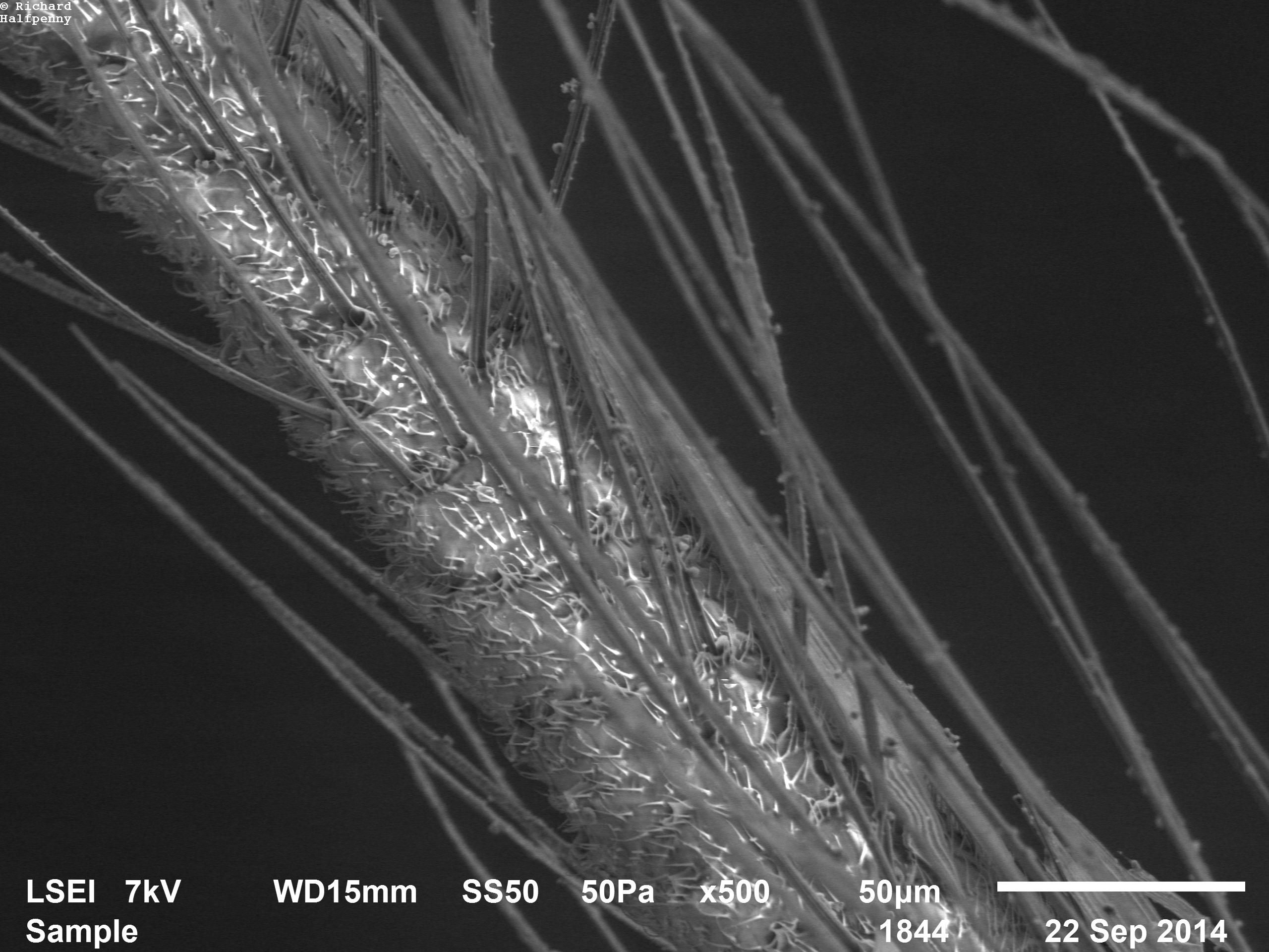
These first 3 images show the complexity, and beauty, of the antennae of this male mosquito. I particularly like the way that new detail is revealed at each new magnification level. From an entirely non-scientific point of view, I find these hugely satisfying aesthetically
Male Culex pipiens antennae. Zoom level 1
Male Culex pipiens antennae. Zoom level 2
Male Culex pipiens antennae. Zoom level 3
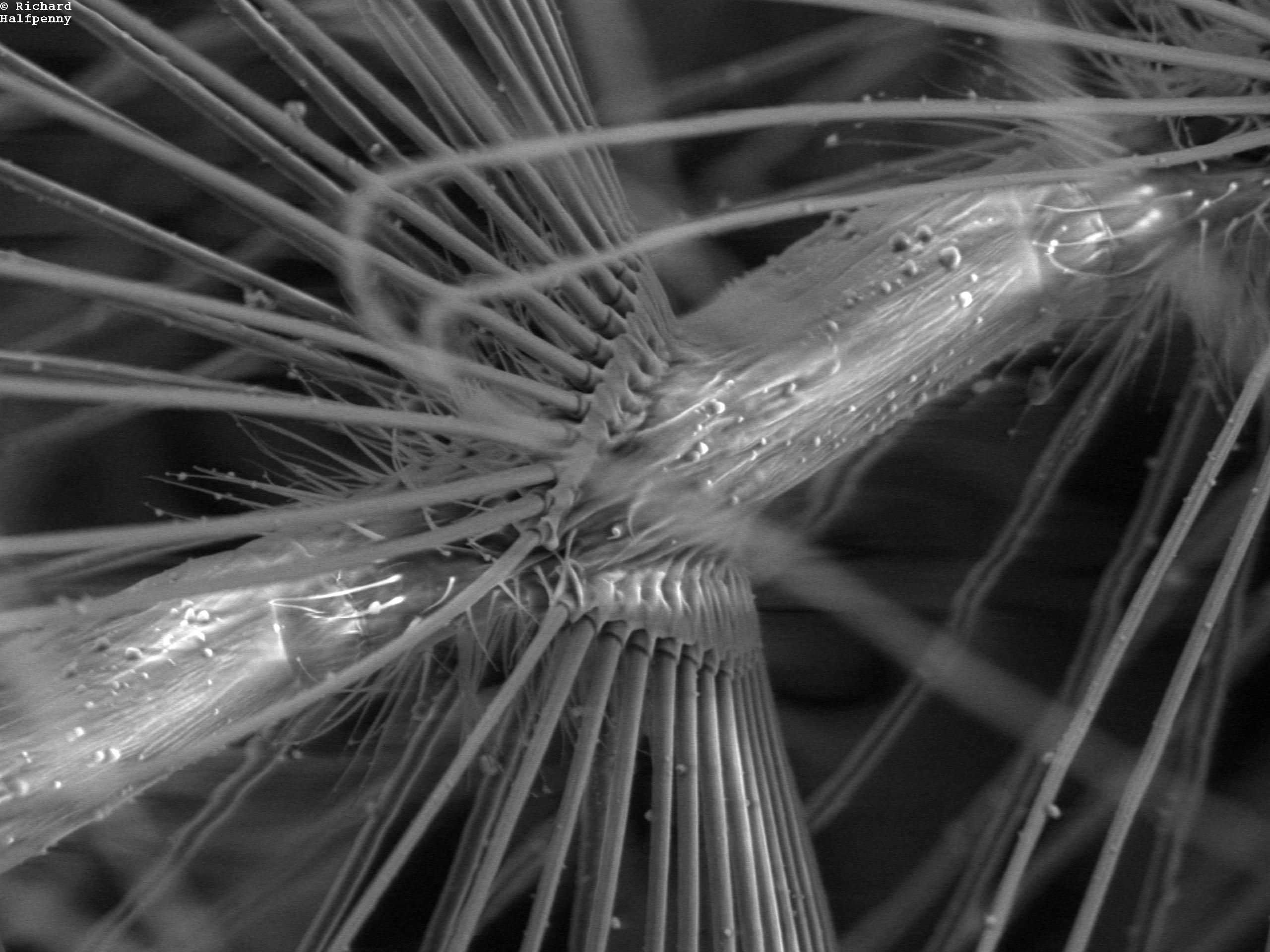
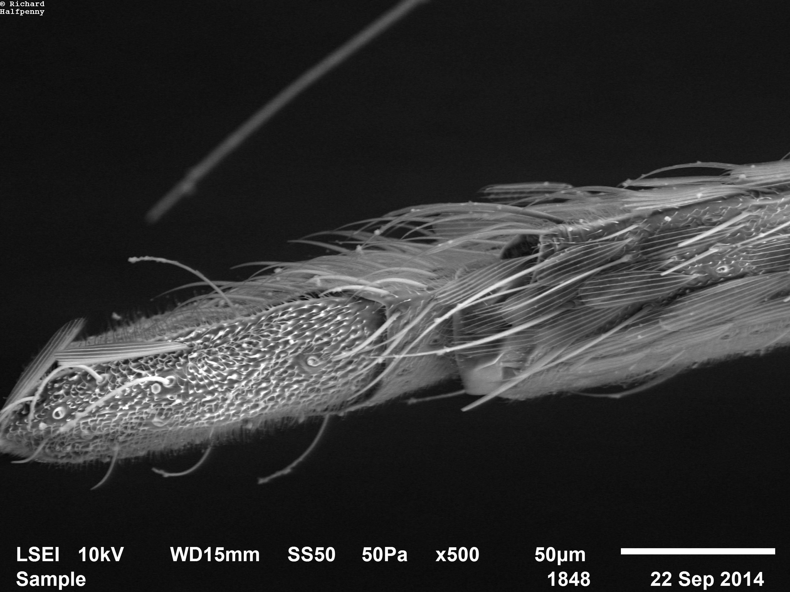
The next 3 images focus on the maxillary palps of the same male Culex pipiens mosquito. Again, starting at low magnification and progressively increasing.

The end of a Male Culex pipiens’ maxillary palp. x100 magnification.
Male Culex pipiens maxillary palp x250 magnification
Male Culex pipiens maxillary palp x500 magnification. (specimen is getting quite charged now- hence the “glowing” hairs)
And last but not least, a 3 image series of the end of the proboscis of the same Culex pipiens male.
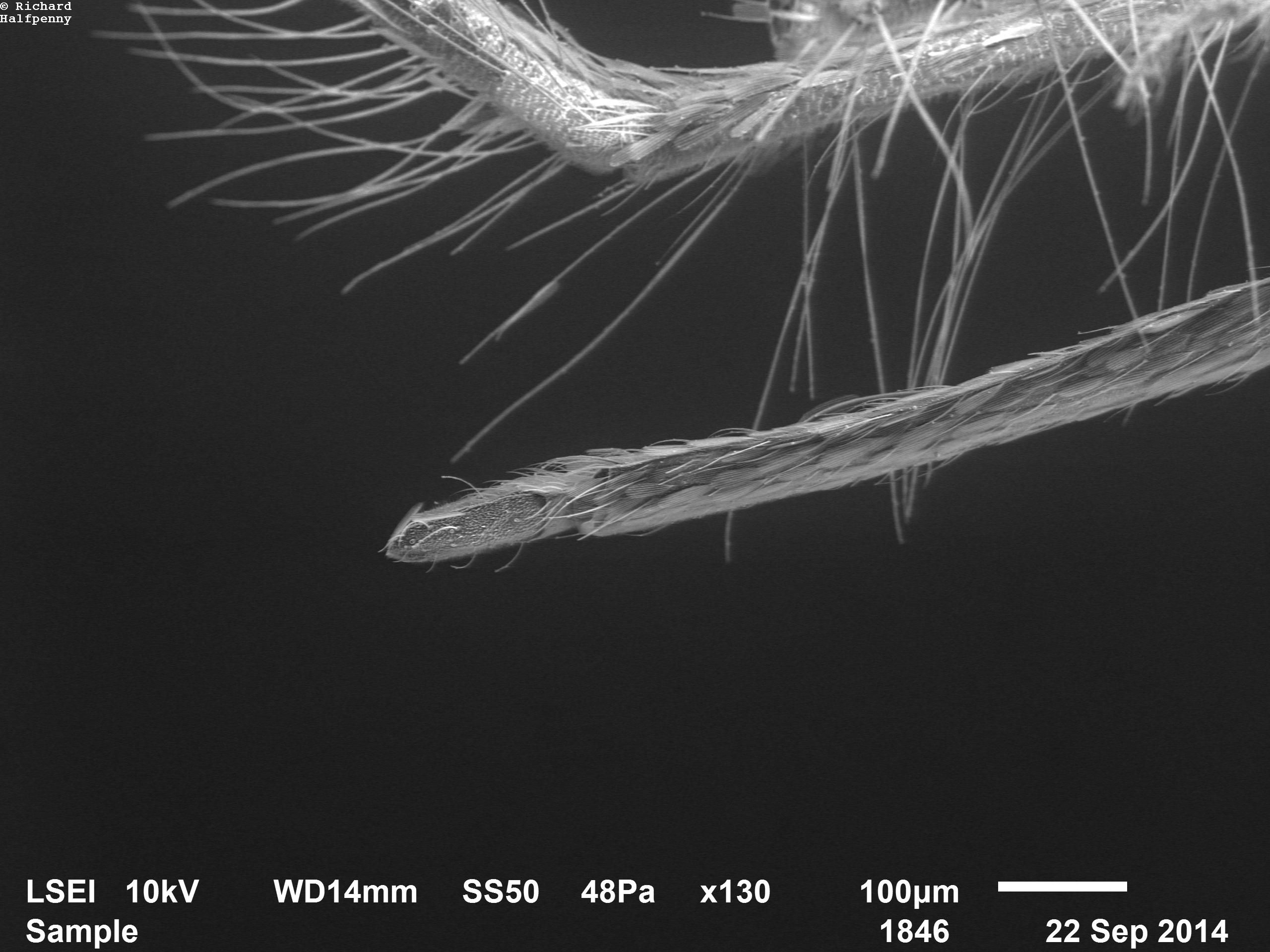
Male Culex pipiens proboscis x130 magnfication
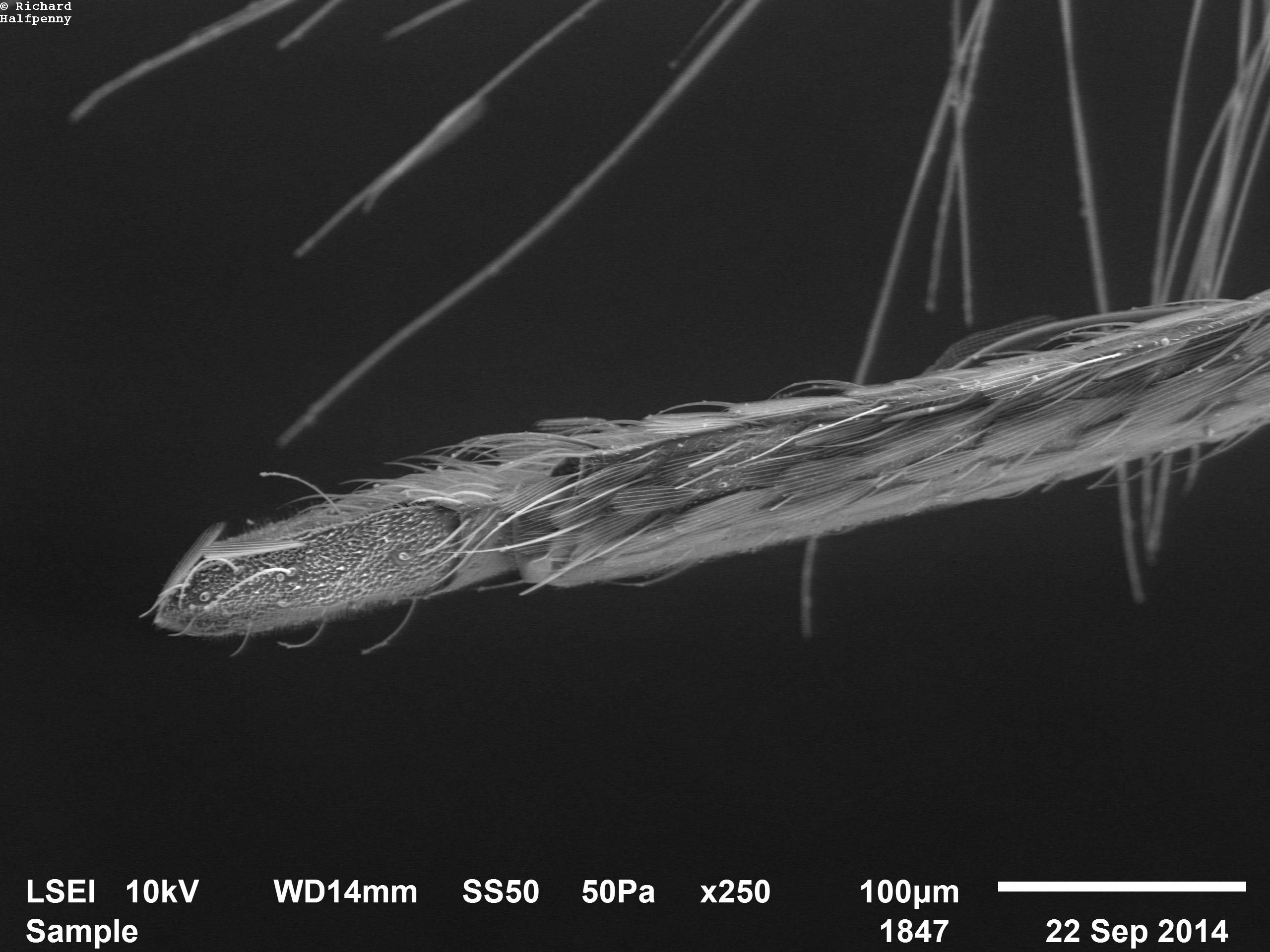
Male Culex pipiens proboscis x250 magnfication
Male Culex pipiens proboscis x500 magnfication
Experienced microscopists please excuse the specimen charging throughout, and the missing scale data on the first 3 images.